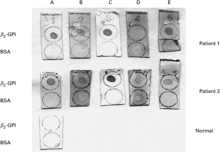
Fig. 2.
Dot blot analysis of two representative serum samples from patients having antibodies (IgG) to β2-GPI before and after inhibition experiments. A, Serum diluted in buffer; B, cardiolipin (CL)-liposomal supernatant; C, CL-liposomal eluant; D, fluid-phase inhibition with 200 μg/ml of β2-GPI; E, fluid-phase inhibition with 100 μg/ml of β2-GPI. Strips were dotted with 3 μg of β2-GPI or bovine serum albumin (BSA). A normal serum is shown as control.

