Fig. 1.

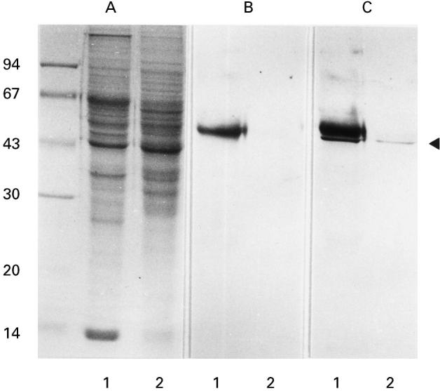
Detection of serum IgG reactive with a 47-kD renal protein in patients with primary MN. A, SDS–PAGE gel stained with coomassie brilliant blue R-250. Numbers on the left indicate molecular mass standards (Pharmacia, Uppsala, Sweden) in kD. Lanes 1 and 2 are isotonic buffer-extractable fractions from human and porcine kidneys, respectively. B, Immunoblot probed only with alkaline phosphatase-conjugated anti-human IgG. A 55-kD immunoreactive protein band in lane 1 is regarded as human IgG heavy chain. C, Immunoblot probed with serum from a patient with primary MN and with alkaline phosphatase-conjugated anti-human IgG. The arrowhead indicates the 47-kD protein bands in the human and porcine renal extracts, which specifically react with the patient's serum IgG.
