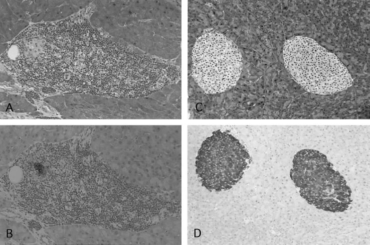
Fig. 3.
Histological analysis of pancreata in NOD mice given IFN-γ-stimulated DC. Sections prepared from the pancreata of 25-week-old control NOD mice (A and B, grade 4; mag. × 100) and 25-week-old NOD mice that had been given IFN-γ-stimulated DC at 4 weeks old (C and D, grade 0; mag. × 80) were stained with haematoxylin and eosin (A,C) or immunohistochemically examined for insulin-producing β cells (B,D).

