
Fig. 10.
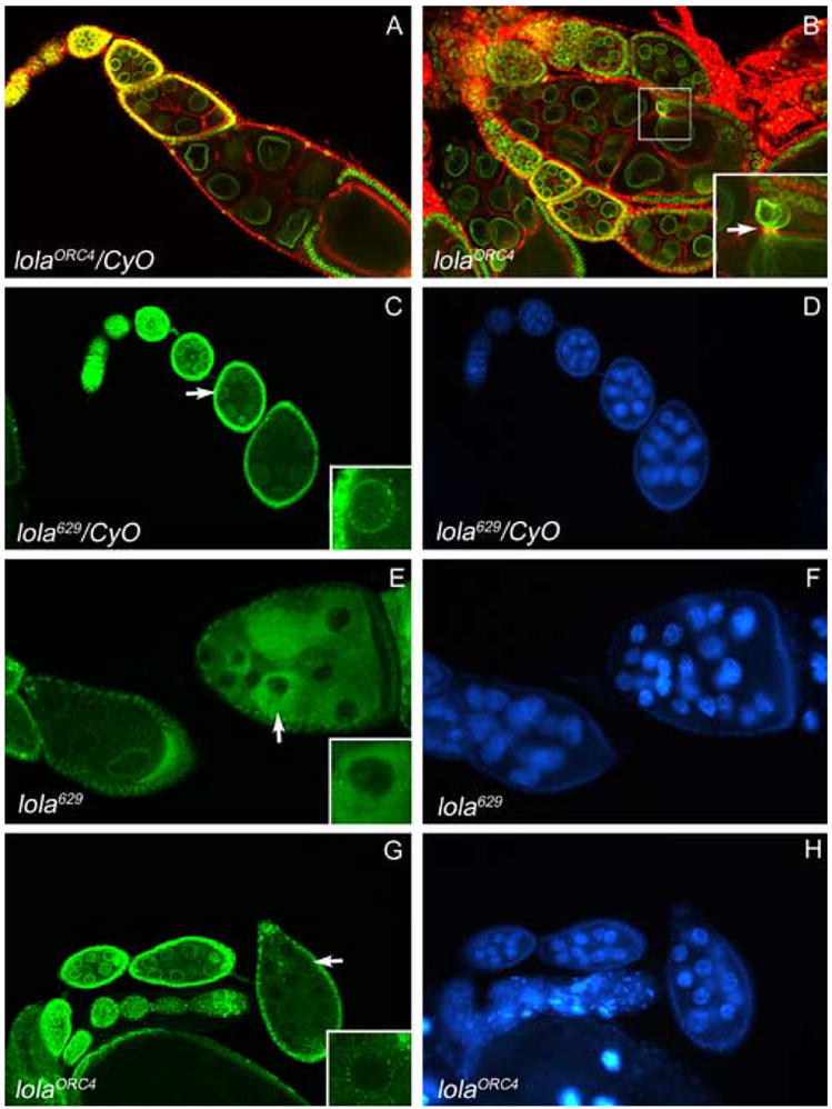
lola629 and lolaORC4 GLCs exhibit abnormal nuclear lamina morphology. Egg chambers in (A–B) were double-labeled with lamin Dm0 ADL84.12 antibody (green) and rhodamine-phalloidin (red). (A) Heterozygous control egg chambers exhibit normal nuclear lamina morphology. (B) A lolaORC4 GLC egg chamber exhibits a protrusion of the nurse cell nuclear lamina through the ring canal (arrow, higher magnification inset). Egg chambers in (C–H) were stained with lamin Dm0 ADL101 antibody (green, C, E, and G) and DAPI to visualize DNA (blue, D, F, and H). (C) Nuclear lamin Dm0 surrounds the nuclei in heterozygous control egg chambers (higher magnification inset of nucleus indicated by arrow). (E and G) lola629 and lolaORC4 GLCs exhibit mislocalization of lamin Dm0 to the cytoplasm ((higher magnification insets of nuclei indicated by arrows). (F and H) No abnormal chromatin morphology is seen in the egg chambers with mislocalized lamin Dm0 (compare with neighboring egg chambers and heterozygous controls in D).