
Fig. 7.
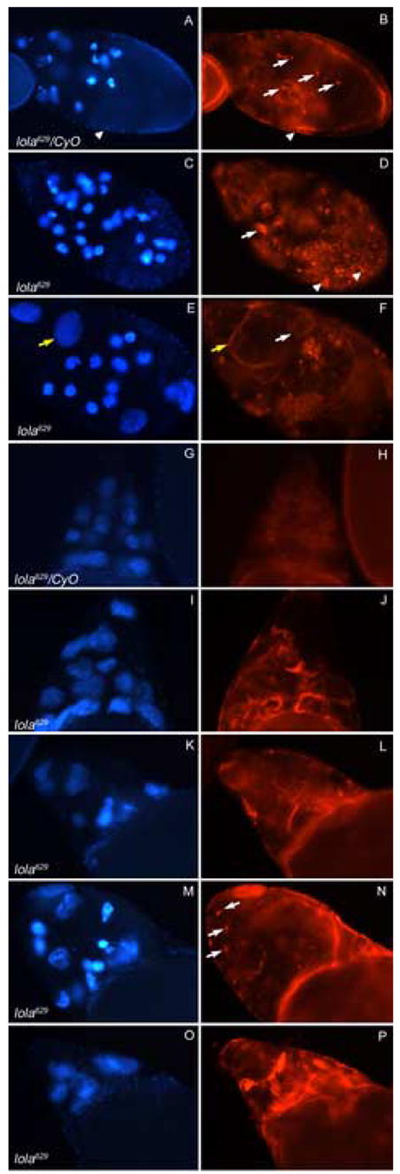
Abnormal actin structure is seen in degenerating lola629 GLC egg chambers. Panels depict DAPI staining (blue) on the left and rhodamine-phalloidin staining (red) on the right of the same egg chambers. All images were taken at the same magnification and rhodamine-phalloidin pictures were taken at the same exposure. (A–B) A heterozygous mid-stage egg chamber undergoing checkpoint PCD, induced through nutrient deprivation, contains condensed nurse cell nuclei, small actin clumps (B, arrows) and follicle cells with cellular membranes still intact (arrowheads). (C–F) lola629 GLC egg chambers undergoing checkpoint PCD, without starvation, contain abnormal actin structures (D and F, white arrows), degenerating follicle cells (D, arrowheads), and uncondensed nurse cell nuclei with intact cellular membranes (E and F, yellow arrows). (G–H) A stage 10B heterozygous egg chamber shows formation of organized actin bundles in all nurse cells. (I–J) A stage 10B egg chamber containing lola629 GLCs exhibits actin bundle formation that is disorganized and not uniform throughout the nurse cells. (K–P) Dumpless egg chambers containing lola629 GLCs continue to show disorganized actin structure. Some contain small actin clumps like those seen during mid-stage PCD (N, arrows).