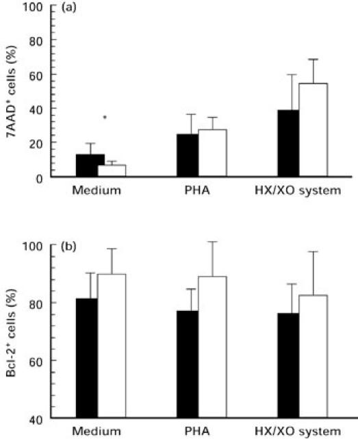
Fig. 5.

Apoptotic measurement after 24-h stimulation with medium alone, phytohaemagglutinin (PHA) 1 μg/ml and hypoxanthine (HX) 1 mm/xanthine oxidase (XO) 10 mU of AT lymphocytes (▪) in comparison with healthy controls (□). Mean values ± s.d. of the (a) 7-aminoactinomycin D (7AAD) incorporation and (b) the number of Bcl-2-expressing cells were detected by flow cytometry; *P < 0.05.
