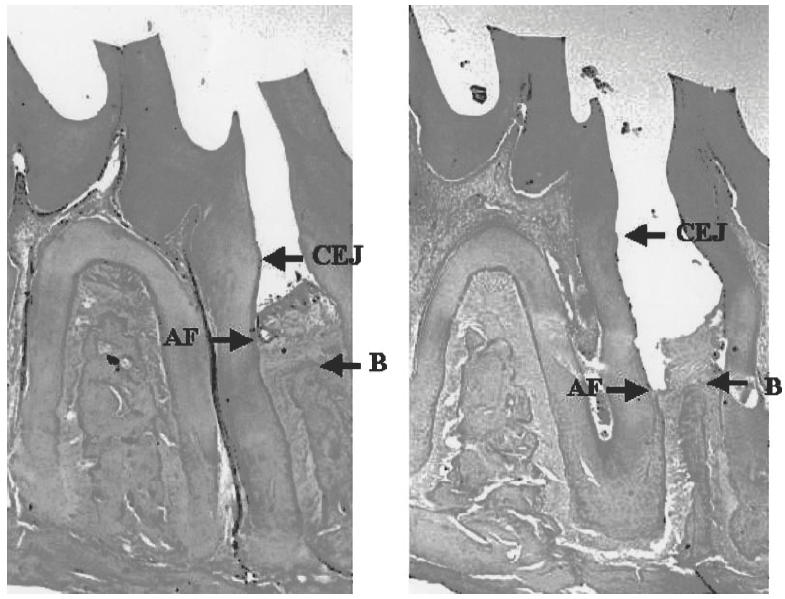
Fig. 1.

Photomicrographs showing histological sections of an experimental tooth in a control Wistar rat injected with 100 μl saline (right), and in a rat prevaccinated with 0·1 mg of SRL172 in 100 μl saline (left). A difference in loss of periodontal attachment fibres (AF) and bone (B) can be observed. CEJ, Cemento–enamel junction; AF, the most coronal attachment fibres; B, alveolar bone crest.
