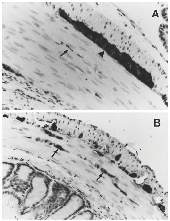
Fig. 2.

Protein gene product (PGP) 9.5-immunoreactive nerve fibres in the muscular layer of colon of an IL-2−/– mouse with chronic colitis (A) and a healthy IL-2+/+ control (B). The arrows point to the positively stained nerve fibres, and the arrowhead indicates a myenteric ganglia. Note that the frequency of the immunoreactive nerves was decreased in the IL-2−/– mouse compared with the IL-2+/+ control. (Original mag. × 400.)
