
Fig. 2.
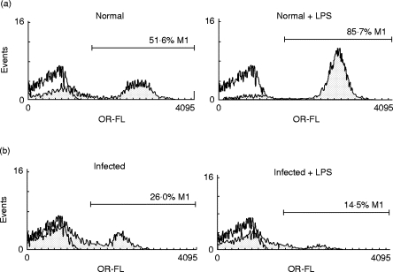
Percentage of CD19+ cells on spleen mononuclear cells (SMC) from Trypanosoma cruzi-infected mice. SMC from normal (a) or 15-day-infected mice (b) were cultured with medium alone (left panels) or LPS (20 μ g/ml) (right panels) during 96 h. The cells were then analysed for CD19+ expression by flow cytometry using PE-labelled anti-mouse CD19 antibodies. Staining with control isotype is shown by open histograms. The relative cell number is plotted against fluorescence intensity.