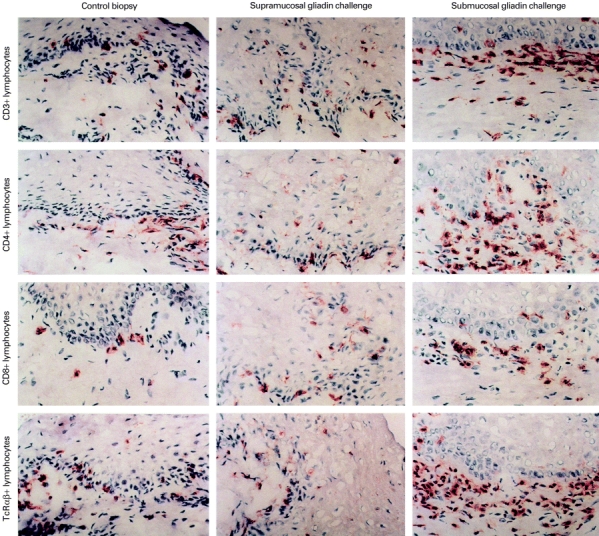
Fig. 1.

(See next page) Immunohistochemical staining of CD3+, CD4+, CD8+ and TCRαβ+ cells in the oral mucosa. A panel of examples of oral mucosal slides in patients with treated coeliac disease before (left panel) and after local oral challenge with gliadin made supramucosally with gliadin powder (middle panel), and submucosally with dissolved gliadin (right panel).
