Fig. 1.
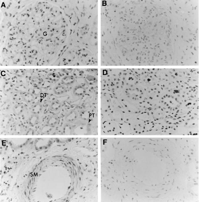
Expression of the C5a receptor by human renal tissue. (A,C,E) Human kidney paraffin-embedded section stained with immunoperoxidase-labelled rabbit anti-C5a receptor antiserum as described in Materials and Methods. (A) Photomicrograph of a section demonstrating the lack of staining within the glomerular tuft (G). (C) Photomicrograph of a kidney section showing C5a receptor expression within the proximal (PT) and distal (DT) renal tubular epithelium. (E) Staining for the C5a receptor within the smooth muscle (SM) of a renal blood vessel. (B,D,F) Background immunostaining using rabbit preimmune serum.

