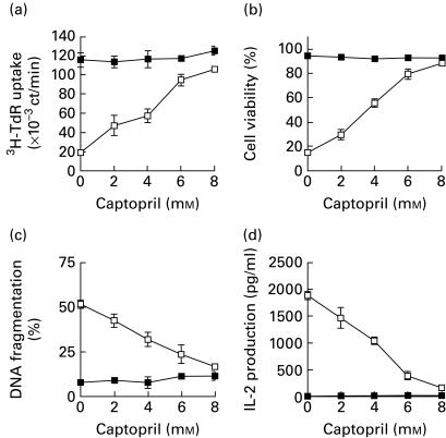
Fig. 1.
Captopril inhibits activation-induced cell death and IL-2 production in T cell hybridoma N3-6-71. (a) Cell proliferation. N3-6-71 cells were cultured in control uncoated 96-well plates or in anti-CD3 antibody-coated 96-well plates (2 × 10⁴ cells/well), in the presence or absence of the indicated concentrations of captopril. After 24 h, the cells were pulsed with 3H-thymidine, and harvested after an additional 2 h. Incorporation of 3H-thymidine into cellular DNA was measured in a scintillation counter. (b) Cell viability. N3-6-71 cells were cultured in control 24-well plates or in anti-CD3 antibody-coated 24-well plates (5 × 10⁵ cells/well), in the presence or absence of the indicated concentrations of captopril. After 50 μl of supernatant were removed from each well for the measurement of IL-2 at 24 h of cultivation, the cells were harvested and cell viability was determined by trypan blue dye exclusion. (c) DNA fragmentation. 3H-thymidine-labelled N3-6-71 cells were cultured in control 96-well plates or in anti-CD3 antibody-coated 96-well plates (2 × 10⁴ cells/well), in the presence or absence of the indicated concentrations of captopril. Twenty-four hours later, the cells were harvested and the percentage of DNA fragmentation was determined as described in Materials and methods. (d) IL-2 production. N3-6-71 cells were cultured as described above for 24 h. Concentrations of IL-2 in the culture supernatant were measured by ELISA. Mean values of triplicates ± s.d. of representative experiments are shown (a, b, c, d). Error bars are less than symbol width when they are not apparent (a, b, c, d). ▪, Control; □, anti-CD3.

