Fig. 2.
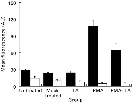
Unstimulated ECV304 cells constitutively expressed moderate levels of ICAM-1 (▪) and MHC-I (□) compared with the isotype antibody controls. Similar levels of expression were detectable in cells treated with dimethyl sulphoxide (DMSO)/methanol or triamcinolone acetonide (TA) alone. Phorbol myristate acetate (PMA)-activated cells had significantly (P = 0·0003) up-regulated ICAM-1 expression after 72 h in culture, an approximately four-fold increase over unstimulated levels of expression. After 24 h initial exposure to PMA cells were additionally exposed to TA (10−6 m) for a further 48 h; treatment of PMA-stimulated cells with TA significantly reduced detectable levels of ICAM-1 expression (PMA versus PMA + TA, P = 0·045). AU, Arbitrary units. MHC-I levels were not significantly modulated by either TA or PMA treatment. Similarly, after 24 h initial exposure to PMA cells were additionally exposed to TA (10−6 m) for a further 48 h; treatment of PMA-stimulated cells with TA did not significantly modulate levels of MHC-I expression.

