Fig. 4.
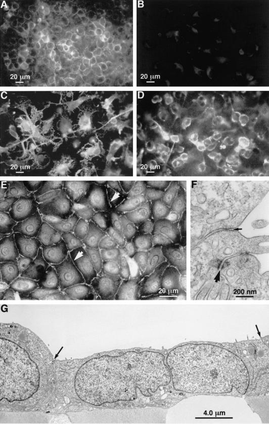
(A) On coverslips, resting ECV304 cells showed cell membrane-localized ICAM-1 immunostaining. (B) On coverslips staining with isotype control (non-immune immunoglobulin) revealed insignificant levels of non-specific binding to ECV304 cells. (C) On coverslips, phorbol myristate acetate (PMA) stimulation promoted hypertrophy and the formation of long processes in ECV304 cells was associated with more intense ICAM-1 expression at the plasma membrane. (D) On coverslips, subsequent treatment of PMA-stimulated cells with triamcinolone acetonide (TA) resulted in condensation of the ECV304 cell morphology associated with smaller cell size and reductions in processes. (E) On coverslips, ZO-1 immunofluorescent staining revealed the ECV304 cells to form an integrated monolayer with high levels of immunoreactivity expressed at the cell margins and occasional punctate accumulations of staining (arrowheads). (F) A high power electron micrograph showing details of a zonule occludentes junction (small arrow) and a maculae adherens (large arrow). (G) An electron micrograph illustrating that ECV304 cells form a continuous layer with apposing cells joined by both desmosomes (maculae adherens) and tight junctions (ZO). Both types of junction occurred at the apices of the cells (arrows indicate cell boundaries).

