
Fig. 1.
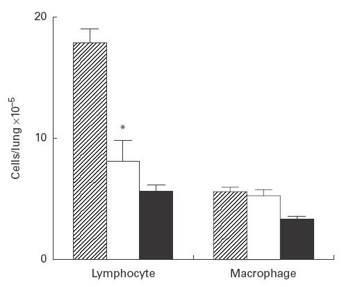
Effects of anti-tumour necrosis factor-alpha (TNF-α) antibody on lymphocyte and macrophage counts in the lung. Pulmonary intraparenchymal lymphocytes and macrophages were examined. The total number of cells was counted, and each population was identified based on morphological analysis of at least 300 cells stained by May–Giemsa staining. Cell numbers in the lung obtained from control (hatched), mice treated with anti-TNF-α MoAb (□) and uninfected (intubated with sterile tubes) mice (▪). Each bar represents the mean ±s.d. of five animals. *P < 0·01 compared with control.