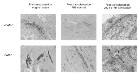
Fig. 5.

Representative samples of the original and grafted synovial tissue, pre- and post-transplantation. Transplants were injected intragraft with PBS (control) or with TNF-α (200 ng/graft). Cryostat sections (10 μm) were stained for anti-human intercellular adhesion molecule-1 (ICAM-1) and vascular cell adhesion molecule-1 (VCAM-1) using standard immunoperoxidase technique. The intensity of staining was scored using an arbitrary scale shown graphically in Fig. 6. M, Murine tissue; H, human tissue; I, interface.
