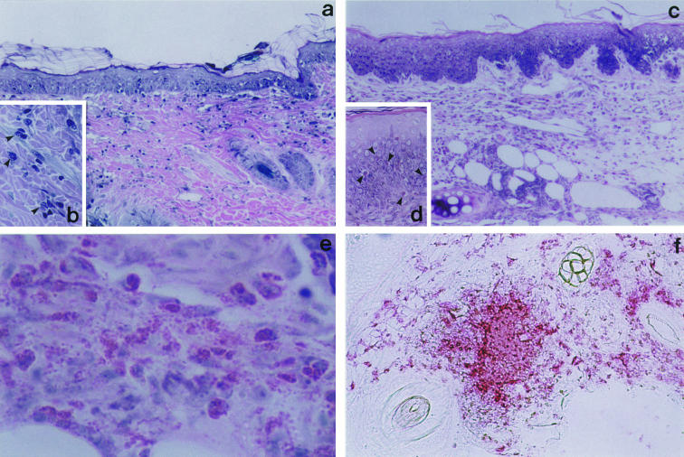
Fig. 1.
Microscopic features of skin reaction in guinea-pig contact sensitivity.Guinea-pigs were sensitized with DNCB and challenged on the dorsal skin or ear lobe on day 14. Excised skin specimens were stained with 10% Giemsa as described in Materials and methods. (a) Dorsal skin. The epidermis shows focal intercellular oedema with mononuclear cell infiltrate (spongiosis). Cellular infiltrate in the upper dermis consists of mononuclear cells, numerous basophils, and a few eosinophils. (b) High magnification view of basophils in the dermis (arrow head). (c) Ear lobe. The epidermis shows spongiosis. The entire dermis contains a remarkable number of eosinophils and mononuclear cells (d) Eosinophils have also infiltrated the epidermis (arrow head). (e) Prominent degranulation of eosinophilic granules. (f) Immunohistochemical staining for MBP in the challenged ear lobe. Numerous cells are positive for MBP and the extracellular deposition of MBP in the dermis is extensive.

