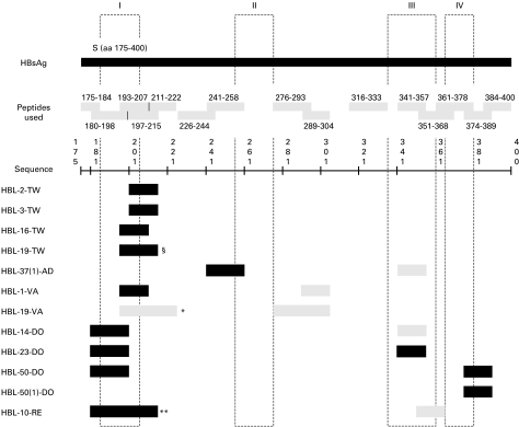
Fig. 1.
Peptide restriction pattern of HBsAg-specific T cell lines. The columns I, II, III and IV represent the four transmembrane regions of the HBsAg, as predicted by Stirk et al. [43]. Peptides that are recognized by HBL-19-TW in §: 193–207 and 197–215; by HBL-19-VA in *: 193–207 and 211–222; by HBL-10-RE in **: 180–198, 193–207, and 197–215. Dark shading refers to a strong peptide recognition and light shading refers to a weak peptide recognition.

