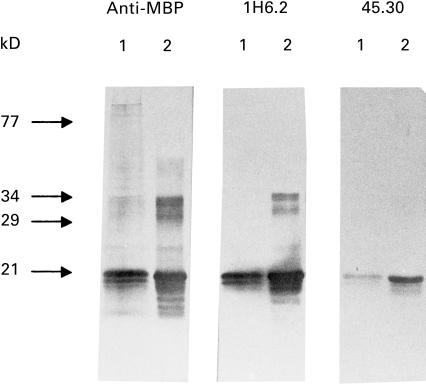
Fig. 2.
Western blot identification of human (lane 1) and bovine (lane 2) MBP using 1H6.2 and 45.30 MoAbs. To compare the reactivity of 1H6.2 and 45.30, a commercially available anti-MBP MoAb was used (anti-MBP recognizing epitope 84-89; Serotec, Oxford, UK). The typical major 18·5-kD MBP band is recognized by both 1H6.2 and 45.30 MoAbs. MBP has a reduced electrophoretic mobility due to its positive charge and runs at approximately the 21 kD level under these experimental conditions [24,25].

