Fig. 2.
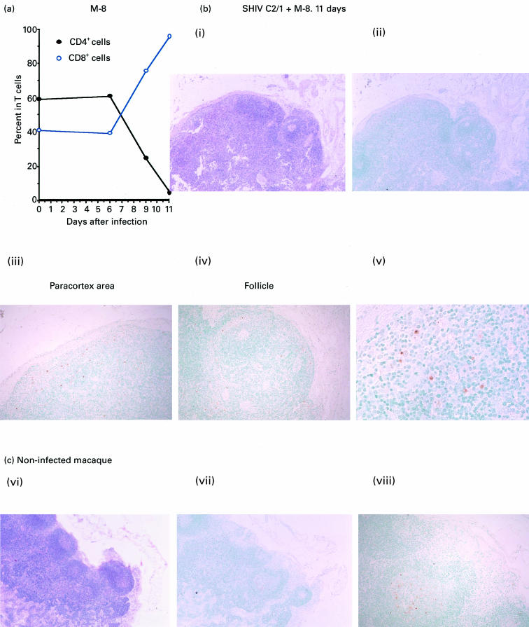
Histological analysis of TdT+ apoptotic cells by the TUNEL method in a submandibular lymph node from an SHIV-C2/1-infected macaque at day 11 post-inoculation. (a) Changes in percentage of either CD4+ or CD8+ cells in T lymphocytes in one representative monkey. (b) Detection of TdT+ cells in a lymph node from an SHIV-C2/1-infected macaque at day 11 post-inoculation. Both Bi and Bii show images of a section of lymph node, Biii shows a magnified image in the paracortex area, and Biv and Bv show magnified images of follicles in the lymph node. (c) TUNEL staining of a submandibular lymph node obtained from an uninfected control animal. Haematoxylin and eosin (Bi and Cvi, mag. × 40), TUNEL (Bii, mag. × 40; Biii and Biv, mag. × 100; Bv, mag. × 400; Cvii, mag. × 40, and Cviii, mag. × 100) staining procedures were used.

