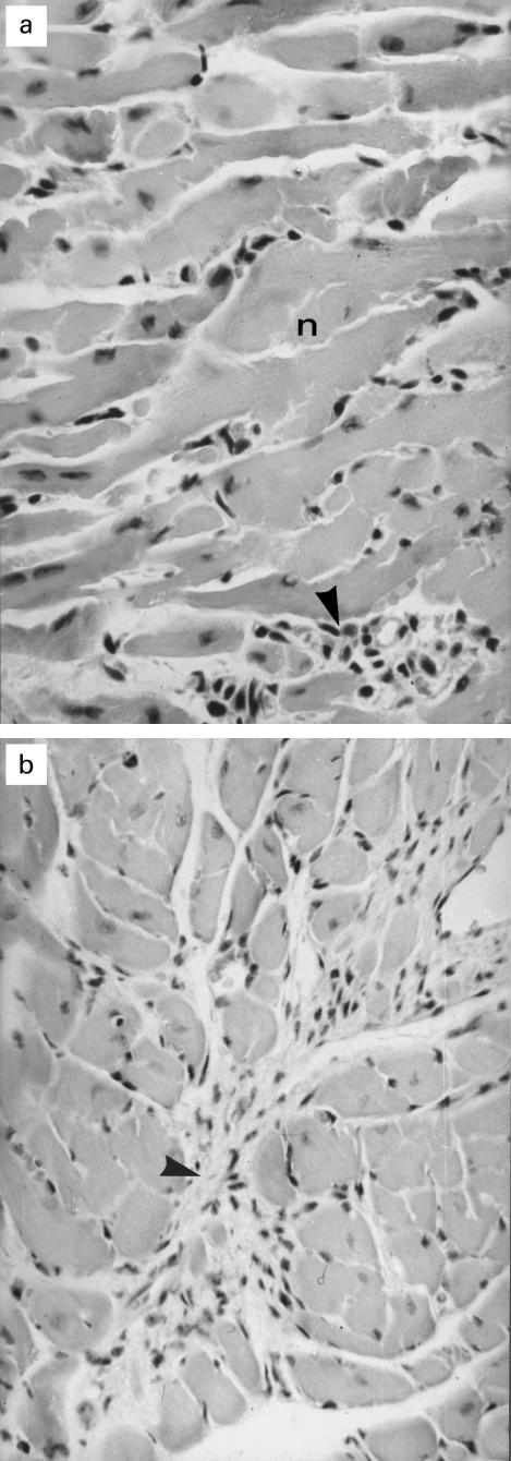
Fig. 1.
Histopathology of hearts of coxsackievirus B3 (CVB3)-infected mice. (a) Myocarditis due to CVB3 infection showing myocytic necrosis (n) and lymphohistiocytic infiltration (arrowhead); mice, 2 weeks p.i., H–E staining; primary mag. × 200. (b) Marked diffuse interstitial fibrosis including mesenchymal proliferation (arrowhead) in the heard of a mouse 13 weeks p.i.; H–E staining; primary mag. × 100.

