
Fig. 1.
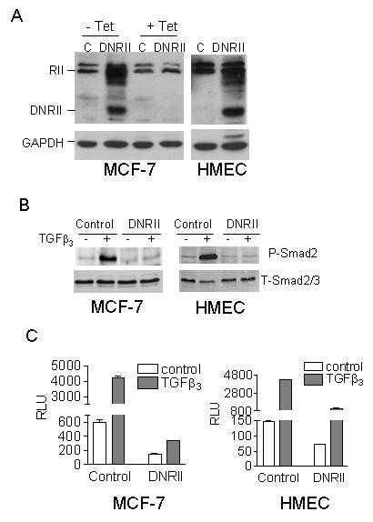
Blockade of TGFβ signaling in MCF-7 and HMEC cells by the expression of a TGFβ DNRII. (A) The expression of DNRII was detected with Western blotting using the lysate of control (C) and DNRII-transfected cells. Since the MCF-7 cells were transfected with a tetracycline (Tet)-suppressible DNRII expression system, they were treated with or without Tet at 1 μg/ml for 5 days before harvested for the Western blotting analysis. GAPDH was blotted to indicate equal sample loading. (B) Exponentially growing cultures of the control and DNRII-transfected MCF-7 and HMEC cells were treated with 2.0 ng/ml of TGFβ3 for 24 h. Cell lysates were collected afterward and Western Blotting was performed for phosphorylated Smad2 (P-Smad2) and total Smad2/3 (T-Smad2/3) as described in Materials and Methods. (C) Control and DNRII-transfected MCF-7 and HMEC cells were transiently co-transfected with a TGFβ-responsive promoter-luciferase construct (p3TP-Lux for MCF-7 cells or pSBE4-Luc for HMECs) and a β-gal expression construct. The transfected cells were treated with or without 2.0 ng/ml of TGFβ3. The activity of luciferase and β-gal in the cell lysates was measured 24h later. The β-gal-normalized luciferase activity (RLU) was plotted. The data represent the means ± SEM from triplicate transfections.