
Fig. 2.
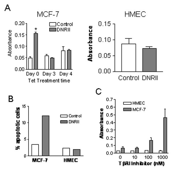
Effect of the blockade of autocrine TGFβ signaling on cell apoptosis. (A) MCF-7 control and DNRII cells were plated in 100-mm dishes at 500,000 cells per dish. Cells were treated with or without tetracycline (0.5 μg/ml), two days after plating, for the indicated time period. HMEC control or DNRII cells were plated at 250,000 cells per 60 mm dish and cultured for 7 days. Apoptosis was measured with Cell Death Detection ELISA using 30,000 cells from each well per manufacturer's instructions. The results plotted represent the means ± SEM from triplicate wells. The treatment with an asterisk “*” denotes a significant (P<0.05) difference from other treatments with one-way ANOVA followed with Newman-Keuls Multiple Comparison Test. (B) The control and DNRII cells of MCF-7 and HMEC were cultured in T-25 flasks and harvested at exponential growth phase. The cells were detached with a trypsin solution and stained with Annexin V-FITC and propidium iodide. The stained cells were analyzed with a flow cytometer. Percentage of apoptotic cells with positive staining by Annexin V-FITC and negative staining by PI is plotted. (C) Parental MCF-7 were plated at 0.5×10⁶ cells per 60-mm dish in McCoy's 5A medium with 1% FBS. hTERT-immortalized HMEC cells were plated at 0.2×10⁶ cells per 60-mm dish in the serum-free MEGM medium. The cells were treated with various concentrations of a TGFβ kinase inhibitor (Calbiochem) for 5 days after plating. The cells were then harvested for the apoptosis ELISA and the results plotted represent the means ± SEM from triplicate wells.