
Fig. 5.
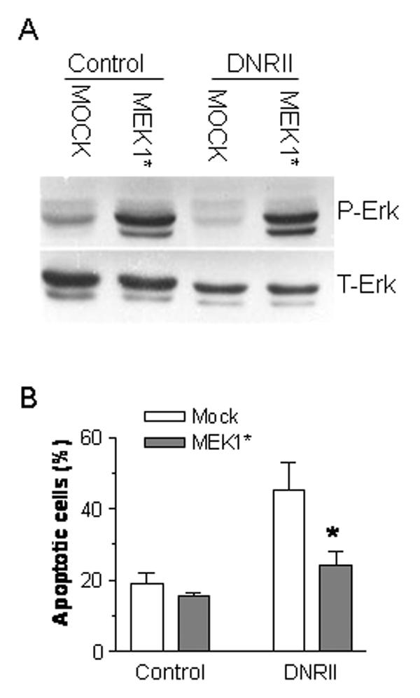
Inhibition of DNRII-induced apoptosis by the activation of Erk in MCF-7 cells. MCF-7 control and DNRII cells were plated at 500,000 cells per 60mm dish and cultured for 24 hr. The cells were co-transfected with a constitutively active MEK1 (MEK1*) expression plasmid and a red fluorescent protein expression vector pDsRed1-N1 (Clontech). After 48 hr, the cells were stained with Annexin V-FITC and the apoptotic cells stained with Annexin V-FITC and also expressing the red fluorescent protein were counted with a flow cytometer. The results plotted in Panel B represent the means ± SEM from three independent transfections. The cells recovered from flowcytometry were used for Western blotting for p-Erk and T-Erk as shown in Panel A.