
Fig. 2. Germline cyst development in cuff mutants.
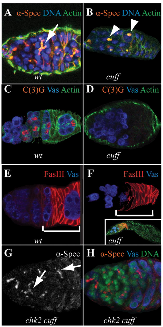
(A, B) Using an antibody against α-Spectrin (red), we always observed highly branched fusomes (arrow) in wild type germaria in the anterior regions (A). In contrast, in cuff mutant germaria, many of the cysts fail to divide normally and form branched fusomes, instead often retaining a round spectrosome-like fusome (arrowheads) (B).
(C, D) Using an antibody against C(3)G (red), we monitored meiotic progression in wild type and cuff mutant backgrounds. In wild type germaria, we consistently observed multiple cysts initiating meiosis (C); while in cuff mutant background, although germ cells are present (as marked by Vas staining in blue), the cysts lack C(3)G staining, indicating an early arrest (D).
(E, F) In wild type germaria, developing germline cysts migrate to the posterior of the germaria (E), and subsequently bud off, forming an egg chamber. In cuff mutants, this process is disrupted. In corresponding regions marked by the bracket (region 2b and 3 as marked by FasIII staining in red), there are few if any normal looking cysts in cuff mutant germaria (F).
(G, H) The defects in cyst development of cuff mutants is partially suppressed by a mutation in chk2. In chk2 cuff double mutants, we observed many highly branched fusomes (arrows in G), and females also lay many more eggs than the corresponding cuff mutant.
All flies were growing under optimal nutritional conditions. Cuff heterozygous females were used as wild type control, while cuff WM25/KG0595 and chk2 cuff WM25females were used for phenotypic analysis.