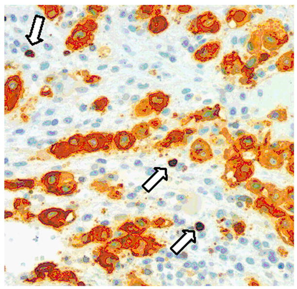
Fig. 1.

Immunohistochemistry of HLA-G and Ki-67 in an exaggerated placental site from an endometrial curettage specimen. HLA-G immunoreactivity is located in the cell membrane and cytoplasm while Ki-67 immunoreactivity is located exclusively in the nuclei. The HLA-G positive intermediate trophoblastic cells do not demonstrate Ki-67 nuclear staining. Therefore, the finding is consistent with an exaggerated placental site, a benign and physiological process during pregnancy. There are scattered Ki-67 nuclei in the filed, and they represent proliferating T-cells or NK-cells (arrows). Without HLA-G immunostaining, it would be difficult to determine the cell type (trophoblastic vs. non-trophoblastic) of the Ki-67 labeled cells.
