
Figure 5.
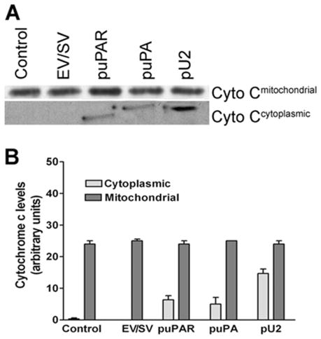
SNB19 cells were transfected with mock, empty vector (EV)/scrambled vector (SV), puPA, puPAR and pU2 plasmid constructs. After 48 h, cells were collected and mitochondrial and cytoplasmic fractions separated as described in Materials and Methods. The mitochondrial fraction was lysed and loaded onto SDS-PAGE gels and after electrophoresis, western blotted and immunoprobed for cytochrome c levels. Cytoplasmic cytochrome c levels, which indicate mitochondrial membrane permeability, were determined (A). Cytoplasmic and mitochondrial cytochrome c levels were quantified and expressed as arbitrary units (B). The values shown are mean ±SD from four different experiments (p<0.01)