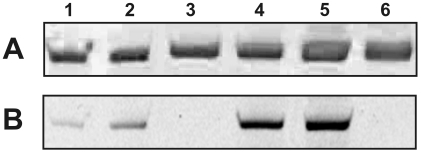
Fig. 1.
Electrophoretic analysis by non-denaturing 5% PAGE of complexes of GTP-TR and α2M* formed at 37°C for 18h (A) Coomassie brilliant blue and (B) fluorescence imaging. The lanes are as follows: lane 1 and 2, proteolytically activated α2M* with GTP-TR; lane 3, proteolytically activated α2M*; lane 4 and 5, non-proteolytically activated α2M* with GTP-TR; lane 6, non-proteolytically activated α2M*.

