
Figure 2.
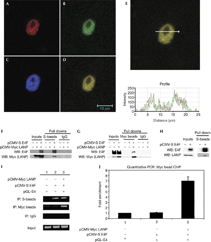
LANP and E4F interact in mammalian cells. (A–E) Confocal immunofluorescence of HeLa cells showing that endogenous LANP and E4F colocalize. Stained for (A) E4F, (B) LANP, (C) DAPI (nuclear), and (D) A and B merged. (E) Fluorescence intensity profile over a random cross-section of the merged image in (D) shows a correlation of intensities of fluorophores used to visualize E4F and LANP. (F,G) Co-precipitation of S-tagged E4F and Myc-tagged LANP in HeLa cells. Western blots (WB) showing the proteins in the immunoprecipitates. (H) In N2A cells, endogenous LANP is precipitated with S-beads when S-tagged E4F is expressed. (I) Chromatin immunoprecipitation (IP) showing that LANP–Myc and S-E4F occupy E4 promoter. (J) Real-time PCR to quantify the amount of pGL-E4 immunoprecipitated with LANP. DAPI, 4,6-diamidino-2-phenylindole; LANP, leucine-rich acidic nuclear protein.