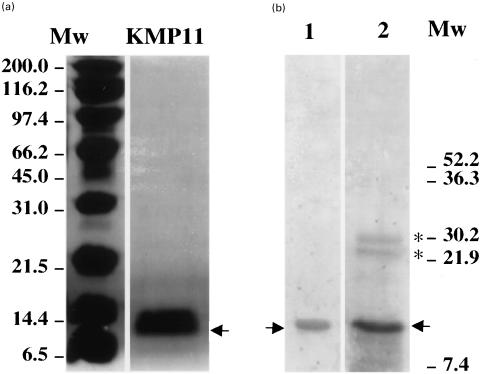
Fig. 1.
SDS-polyacrilamide gel electrophoresis of T. cruzi KMP11 purified recombinant protein. (a) Coomassie blue staining. Lane KMP11, shows the KMP11 recombinant protein purified by Ni2+ -NTA-agarose affinity column (Quiagen). (b) Western blot analysis. Lane 1, filter probed with a pool of five chagasic sera. Lane 2, filter probed with the anti-KMP11 antibody purified by affinity chromatography from a rabbit hyperimmune sera [10]. Mw, molecular weight markers. The arrow indicates the localization of the KMP11 protein. The asterisks show the dimeric and trimeric forms of the KMP11 protein.

