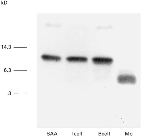
Fig. 2.
SAA degradation by peripheral blood monocytes. CD3+ T cells, CD20+ B cells and CD14+ monocytes (Mo) were isolated from peripheral blood using magnetic beads. These isolated cells (1 × 10⁵/well) were incubated with SAA (10 µg/ml) for 3 h. Culture supernatants were subjected to anti-SAA immunoblot analysis. SAA indicated nondegradated SAA standard. A representative example of two independent experiments showing similar results.

