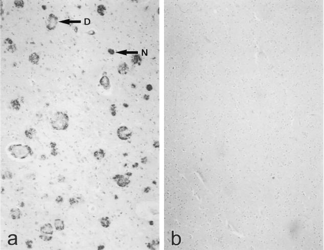
Fig. 2.
Immunohistochemical staining of 4-μm sections of paraffin-embedded, formalin-fixed human Alzheimer's disease (AD) and control temporal lobe using MoAb 3B9. Staining of neuritic plaques (N) and diffuse plaques (D) is apparent in tissue from an AD patient (a) but not in tissue from a non-demented control individual (b).

