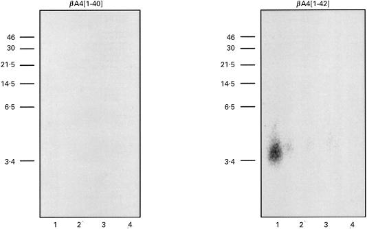
Fig. 6.
Immunoprecipitation of 125I-labelled βA4 using MoAb 3B9 (lane 1) showing recognition of βA4[1–42] but not βA4[1–40]. Samples include: lane 1, 125I-βA4[1–40] or [1–42], MoAb 3B9, RAM-IgG, Pansorbin cells; lane 2, 125I-βA4[1–40] or [1–42], isotype control ET-1 MoAb, RAM-IgG, Pansorbin cells; lane 3, 125I-βA4[1–40] or [1–42] and Pansorbin cells; lane 4, 125I-βA4[1–40] or [1–42], RAM-IgG and Pansorbin cells. Immunoprecipitation performed as described above.

