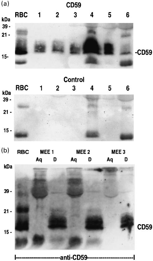
Fig. 4.
(a) Western blotting analysis of CD59 in six different MEE samples. The MEE samples were diluted in a non-reducing sample buffer and run on a 15% SDS-PAGE gel, transferred to a nitrocellulose membrane and immunostained with the BRIC 229 (anti-CD59) MoAb. As a positive control CD59 prepared from a lysate of human red blood cells (RBC) was used. CD59 can be detected on all samples shown. In the control the anti-CD59 antibody was omitted. Lanes 1–6 represent samples 6, 12, 17, 18, 19 and 20 in Table 2, respectively. (b) Western blotting analysis of three pools of MEE samples after Triton X-114 phase separation. Aqueous (Aq) and detergent (D) phases from the TX-114 phase separation were immunoblotted using the BRIC 229 anti-CD mAb. A positive control was prepared from a red blood cell lysate (RBC). CD59 (mw 18 kDa) can be detected in the RBC, and only in the detergent phases (D) of the pooled MEE samples at 19–21 kDa.

