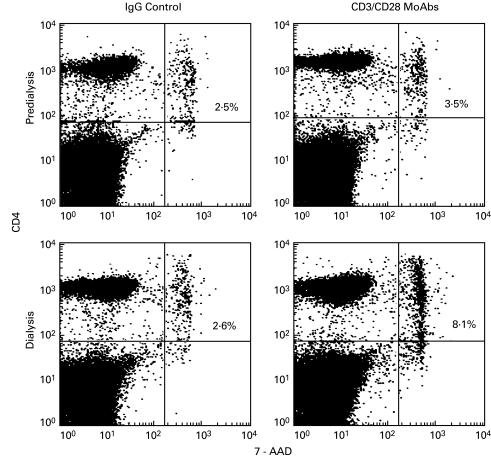
Fig. 5.
Antibody-induced T cell death. PBMC from a dialysis patient were cultured for 18 h with IgG control or anti-CD3/CD28 antibodies, and stained with fluorochrome-conjugated anti-CD4 and anti7-AAD monoclonal antibodies. Data from one representative experiment show the percentage flow cytometry analysis of CD4+ T cells that underwent activation-induced cell death (7-AAD staining). The percentage of cells stained by 7-AAD dye is indicated in the upper right quadrant.

