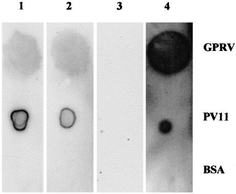
Fig. 5.
Dot blot analysis showing the specific binding of fusion proteins to GPRV and PV11·1µg GPRV, PV11 and BSA were dotted onto PVDF membrane. These were detected with III-20-Fc (lane 1), IV-67-Fc (lane 2), Fc (lane 3, negative control) and HRIG (lane 4, positive control) followed by HRP conjugated anti-human IgG. The blots were developed using chemiluminescence.

