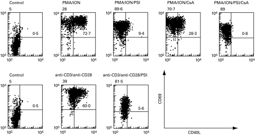
Fig. 2.
PSI and CsA inhibit CD40L surface expression on activated T cells. T cells were stimulated with PMA (50 ng/ml) and ionomycin (1 µm) for 6 h (upper panel) or with immobilized anti-CD3 MoAb (UCHT1) and soluble anti-CD28 MoAb (9·3) (100 ng/ml) for 24 h (lower panel). PSI (200 µm) and/or CsA (400 ng/ml) were added as indicated. After the respective incubation time, the surface levels of CD40L and CD69 on the T cells were analysed by flow cytometry, following immunostaining with anti-CD40L MoAb and anti-CD69 MoAb. Dot plots of reactivity with FITC-conjugated anti-CD40L (x-axis) versus PE-conjugated anti-CD69 (y-axis) are shown.

