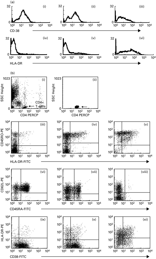
Fig. 1.
(a) RFI of CD38 and HLA-DR CD4+ T cells. (i, iv) Asymptomatic patient. (ii, v) Mild-symptomatic patient. (iii, vi) Patient in category (iii). RFI of activation markers, especially HLA-DR, were strongly elevated in symptomatic patients, whereas the RFI of CD38 was not. (b) Analysis of CD4+ T cell subsets. (i, ii) Gating of CD4+ T cells. The acquisition gate was defined using low SSC and high expression of CD4 to exclude monocytes. (iii, vi, ix) Asymptomatic patient. (iv, vii, x) Mild-symptomatic patient. (v, viii, xi) Patient in clinical category (iii). (iii, iv, v) Analysis of memory and memory-activated CD4+ T cells. In HIV-infected children we found a progressive increase in memory and memory-activated T cell numbers correlating with the progression of the immunodeficiency. (iii, iv, v) Analysis of naive CD4+ T cells. Naive cells were defined as cells with bright expression of CD45RA and positive for CD62-L. In HIV-infected children we found a progressive decrease in naive cell numbers and an increase in CD62L cells correlating with the progression of the immunodeficiency. (vi, vii, viii) Analysis of activated CD4+ T cells. In HIV+ children we found elevated levels of CD4+ CD38+ HLA-DR+ cells. Most cells in asymptomatic patients expressed CD38 at low intensities but these cells did not co-express HLA-DR. These cells are mostly naive cells.

