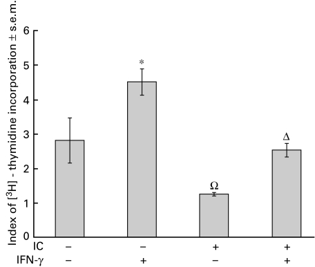
Fig. 6.
Effect of IC on antigen presentation. Adherent PBMC were obtained as indicated in Materials and methods and were incubated with medium or IC (100 µg/ml). After 1 h, the cells were incubated with IFN-γ (240 U/ml) for 24 h. Then, the cells were washed and incubated with non‐adherent PBMC in the absence or presence of Mycobacterium tuberculosis. The index of [3H]-thymidine incorporation was calculated as indicated in Materials and methods. Statistical significance was calculated using the Mann–Whitney test, two-tailed; n = 5. *P < 0·05 significantly different from control. ΩP < 0·03 significantly different from control and IFN-γ-treated cells. ΔP < 0·03 significantly different from IFN-γ-treated cells.

