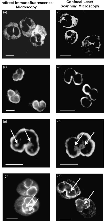
Fig. 1.
Immunofluorescence micrographs of staining patterns produced by different types of antineutrophil antibodies on ethanol-fixed human neutrophils examined by planar and confocal laser scanning indirect immunofluorescence microscopy. Labelling of ethanol-fixed neutrophils by antineutrophil antibodies was detected with FITC-conjugated goat antihuman IgG secondary antibodies. The micrographs in panels a, c, e and g have been visualized by indirect planar immunofluorescence microscopy, the images in panels b, d, f and h were taken by confocal laser scanning microscopy. (a and b) A diffuse cytoplasmic fluorescence, frequently accentuated between the nuclear lobes of the neutrophils, was characteristic of a serum with c-ANCA from a patient with Wegener's granulomatosis. (c and d) A fine homogeneous rim-like staining of the perinuclear cytoplasm was obtained with a serum containing p-ANCA from a patient with microscopic polyangiitis. (e and f) Atypical p-ANCA ‘type A’ gave a broad inhomogeneous rim-like staining of the nuclear periphery along with scattered intranuclear fluorescent foci (arrows). The atypical p-ANCA ‘type A’ were found in the serum of a patient with primary sclerosing cholangitis. (g and h) In addition to the characteristic staining pattern of atypical p-ANCA ‘type A’, atypical p-ANCA ‘type B’ produced a diffuse cytoplasmic fluorescence. The atypical p-ANCA ‘type B’ were detected in a serum from a patient with autoimmune hepatitis. Note that p-ANCA (c,d) can be distinguished from atypical p-ANCA (e–h) in that the atypical p-ANCA show multiple intranuclear fluorescent foci representing stained invaginations of the multilobulated neutrophil nuclear envelope. Size bars indicate 10 µm.

