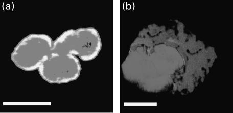
Fig. 4.
Microscopic immunofluorescence patterns of p-ANCA and atypical p-ANCA on formaldehyde-fixed neutrophils with respect to propidium iodide counterstaining. p-ANCA and atypical p-ANCA were detected using FITC-conjugated goat antihuman IgG secondary (dark grey). Propidium iodide counterstaining, was used to label the nucleus (light grey). When both images were optically superimposed, a co-localization of both fluorescence signals was indicated by a bright white staining. The fluorescence patterns were visualized by confocal laser scanning microscopy. (a) Atypical p-ANCA gave a rim-like labelling of the nuclear periphery along with scattered intranuclear fluorescent foci which both colocalized with the propidium iodide staining. (b) p-ANCA showed a diffuse labelling of the cytoplasm which did not overlap with the nuclear propidium iodide staining. Size bars indicate 10 µm.

