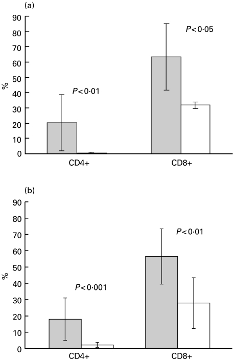
Fig. 1.
(a) CD4+ and CD8+ cells expressing serine esterase (SE) in B–CLL patients and controls. Cytocentrifuged PBMC from patients and controls were stained for SE and CD4+ or CD8+ as described in the Methods. At least 200 CD4+ and CD8+ cells were counted by light microscopy. The values represent mean ± standard deviation. Grey bars = patients: open bars = controls. (b) Perforin-containing CD4+ and CD8+ cells in B‐CLL patients and controls. PBMC stained with anti-CD4 or anti-CD8 MoAbs, fixed, permeabilized and stained with anti-PF MoAbs. Percentages of PF+ cells determined by flow cytometry as described in the Methods. All experiments were carried out in duplicate. The values represent mean ± standard deviation. Grey bars = patients; open bars = controls

