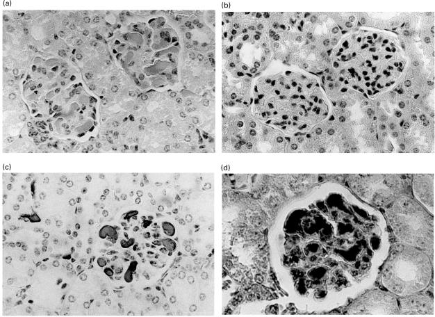
Fig. 1.
Representative haematoxylin and eosin staining of paraffin wax sections from the kidneys of SCID mice implanted with (a) hybridoma cells secreting RH14, showing hyaline thrombi in the glomeruli, (b) control human hybridoma secreting CL24, showing normal kidney morphology. Paraffin wax sections from the kidney of a SCID mouse implanted with RH14 showing the hyaline thrombi stained with antihuman IgG and DAB (c) and PTAH staining of fibrin deposited in the thrombi (d). The magnification of all figures is × 400.

