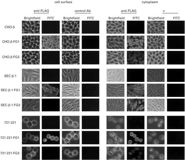
Fig. 5.
Cell surface and cytoplasmic immunohistochemical staining of FG1 and FG3 transfectants. The parental cells and transfectants were stained with anti-FLAG MoAb and isotype control antibody. Bright-field represents the image of each specimen. CHO-β.FG1, CHO transfectant with hβ2m and FG1; CHO-β.FG3, CHO transfectant with hβ2m and FG3; SEC-β.FG1, SEC transfectant with hβ2m and FG1; SEC-β.FG3, SEC transfectant with hβ2m and FG3; 721.221-FG1, 721.221 transfectant with FG1; 721/221-FG3, 721/221 transfectant with FG3.

