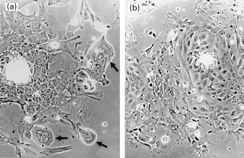
Fig. 2.
Appearance of murine extrahepatic biliary epithelial cells infected by murine cytomegalovirus in vitro and grown for 4 days. Note the large, multi-nucleated, giant cells with smooth contours (arrow, 2a) (×100). Mock-infected MEBEC did not have similar changes over the same period (2b) (×100).

