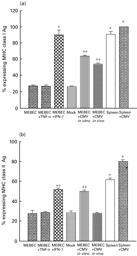
Fig. 4.
Expression of class I antigen (a) and class II antigen (b) by cultured biliary epithelial cells with different culture characteristics by using flow cytometric analysis. MEBEC: cells cultured in growth medium (GM) only; MEBEC + TNF-α: GM supplemented with 100 U/ml TNF-α; MEBEC + INF-γ: GM supplemented with 100 U/ml INF-γ; mock: GM supplemented with supernatant from non-infected mouse embryonic fibroblast cultures; MEBEC + CMV in vitro: cultured biliary epithelial cells in each well exposed to 1·5 × 105 pfu murine cytomegalovirus in GM for 3 days; MEBEC + CMV in vivo: cultured biliary epithelial cells isolated from mice inoculated with 1 × 10⁶ pfu murine cytomegalovirus; spleen: splenocytes from untreated BALB/c mice; spleen + CMV: splenocytes from BALB/c mice inoculated with 1 × 10⁶ pfu murine cytomegalovirus 2 weeks before sacrifice. *P < 0·05; **P < 0·01, when compared with control (cultured in GM only). Results were the average of six separate experiments, error bars represent standard error.

