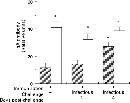
Fig. 1.
ELISA levels of IgA antibodies in sera of immunized C3HeB and BALB/c mice are expressed in units relative to a standard murine anti-Sendai virus antiserum. All values from immunized mice differed (F = 7·9, all P < 0·001) from non-immune mice of the same strain, which generated the background optical density (zero units, not shown). The IgA antibody levels in BALB/c mice were significantly higher than in C3HeB mice (* denotes t > 2·0, P < 0·05 between strains within treatment groups). In C3HeB mice, IgA levels increased significantly by 4 days post-challenge (‡ denotes t > 2·5, P < 0·05 versus other values within the strain), but the IgG levels did not differ significantly from immunized syngeneic mice that were not challenged (not shown).  , C3HeB; □, BALB/c.

