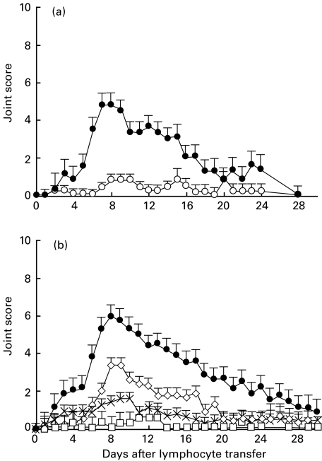
Fig. 4.
Depletion of an activated population abrogates adoptive transfer of polyarthritis by thoracic duct (TD) CD4+ T cells. Purified CD4+ T lymphocytes were prepared from pools of TD lymph from arthritic donor rats by negative selection. They were further depleted of cells expressing activation markers and transferred by intravenous injection to normal syngeneic recipients. MoAbs directed against activation markers were used either singly or in combination or were replaced by the isotype-matched control MoAb (see below). Equal numbers of cells remaining after the secondary depletion with immunomagnetic beads were transferred to recipients in each experiment. The number of cells transferred represented approximately the yield obtained by each procedure from the overnight output of one donor rat and ranged from 5 × 10⁷ to 1 × 10⁸. (a) Transfers of CD4+ T lymphocytes remaining after depletion with a control MoAb (n = 6) or a mixture of MoAbs OX6, OX39, OX26 and OX40 against MHCII, CD25, CD71 and CD134, respectively (n = 5). Joint scores (mean ± SEM) represent the results from three separate experiments. Mixed model analysis showed the curves to be significantly different for days 5–17 (P < 0·0001). •, Control MoAb depleted; ○, anti-MHCII + anti-CD25+ anti-CD71 + anti-CD134 depleted. (b) Transfers of CD4+ T lymphocytes remaining after depletion with either control MoAb IB5(n = 19), MoAb OX39 against CD25 (n = 8), MoAb OX6 against MHC II (n = 12) or a mixture of the MoAbs against MHCII and CD25 (n = 4). •, Control MoAb depleted; ◊, anti-CD25 depleted; ×, anti-MHCII depleted; □, anti-MHCII + anti-CD25 depleted. Data are combined from 10 separate experiments. Comparisons between transfers of cells from each depletion protocol were made using mixed model analysis (see Materials and methods). The course of polyarthritis was significantly different (P < 0·0001) only for the preparations and periods not bearing a common letter (a, b, c) as designated in the following; •, depletion with negative control MoAb, adays 2–30; ◊, depletion of CD25+ cells, bdays 2–5, 20–30, cdays 6–19; ×, depletion of MHC class II+ cells, bdays 2–30; □, depletion of cells expressing CD25 and/or MHC class II,b days 2–30.

