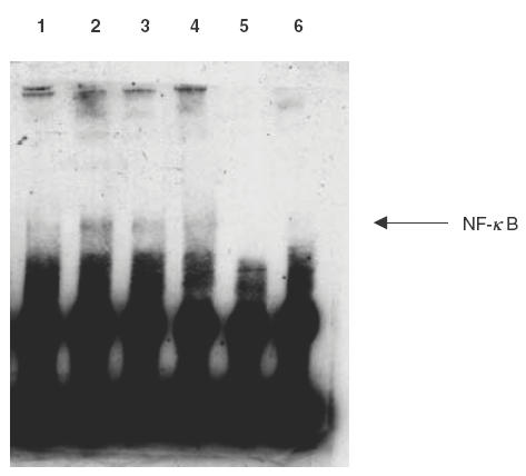
Fig. 3.

EMSA for NF-κB translocation and DNA binding. Eosinophils (5 × 10⁶) were pretreated with or without MG132 (20 μm) or NaSal (20 mm) for 1h followed by stimulation with TNF-α (20 ng/ml) for 12 h. Nuclear protein was isolated, and EMSA was performed with a biotin end-labelled NF-κB oligonucleotide using LightShift™ chemiluminescent EMSA kit (Pierce). Lane 1–4 were as labelled. Lane 5 had only biotinated NF-κB DNA probe without protein added and lane 6 had biotinated NF-κB DNA probe, excess unlabelled NF-kB DNA probe and TNF-treated protein extract. This result is representative of essentially identical results of triplicate experiments. 1: no treatment; 2: TNF (20ng/ml); 3: MG132 (20μm) and TNF (20ng/ml); 4: NaSal (20μm) and TNF (20ng/ml); 5: negative control (biotinylated NF-κB probe only); 6: biotinylated NF-κB DNA probe, TNF-treated protein extract and excess unlabelled NF-κB DNA probe.
