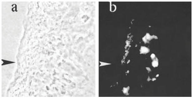
Fig. 1.

Section of gall bladder of a Tnfsf5–/– mouse infected with Cryptosporidium parvum under (a) phase contrast and (b) incident u.v. and green fluorescence filters (both ×100). The arrow marks the serosal surface of the gall bladder, which is thickened as a result of inflammation. Positive staining for TNFα in (b) is localized principally to the submucosa. No green fluorescence was seen in the same tissue stained with an isotype control or uninfected tissue stained for TNFα (not shown).
